Earth sciences, mineral resources, energy and other resources
The earth sciences deal with the physical and chemical processes that affect the earth. Such processes take place on both a macroscopic as well as a microscopic level. A detailed insight into these structures helps us to better understand and interpret the internal processes and developments of the earth and its geological history. Such findings allow, among other things, the optimisation of the mining and extraction of mineral resources and raw materials.
In terms of a sustainable energy industry, the reclamation of raw materials is playing an increasingly important role in the global raw material cycle. In this respect, the exact chemical classification is of vital importance.
JEOL solutions support you in these types of tasks by characterising and analysing solid, liquid and gaseous materials. JEOL is a leader in the innovative energy carriers sector, and supplies unique solutions for determining the concentration and dynamics of lithium and its compounds.
Detailed solutions
- Analysing paper
Analysing paper
As a mass-produced product, it is possible to manufacture paper cost effectively if the ratio of fibres and filler material is optimised without the changing the mechanical and printing properties. In this respect it is important to examine the distribution of fibre and filler material in the paper both during the paper development and production. The exceptionally robust analytic systems from JEOL can be used during challenging routine operation for artifact-free preparation, as well as for the precise morphological and chemical characterisation of cellulose.
Product groups
Solutions
Earth sciences, mineral resources, energy and other resources
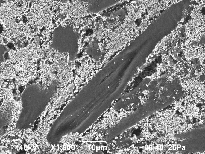 Facing cut through a sheet of paper
Facing cut through a sheet of paper
Source data: JEOL (Germany) GmbH
- Asbestos analysis
Asbestos analysis
Asbestos was used for decades as a fire- and temperature-proof raw and insulation material. Once the health risks were discovered, many laboratories examined potentially asbestos-containing construction materials. JEOL is the only manufacturer to offer the powerful combination of its own electron microscopes and own spectrometers as a complete solution for standards-compliant asbestos analysis.
Product groups
Solutions
Earth sciences, mineral resources, energy and other resources
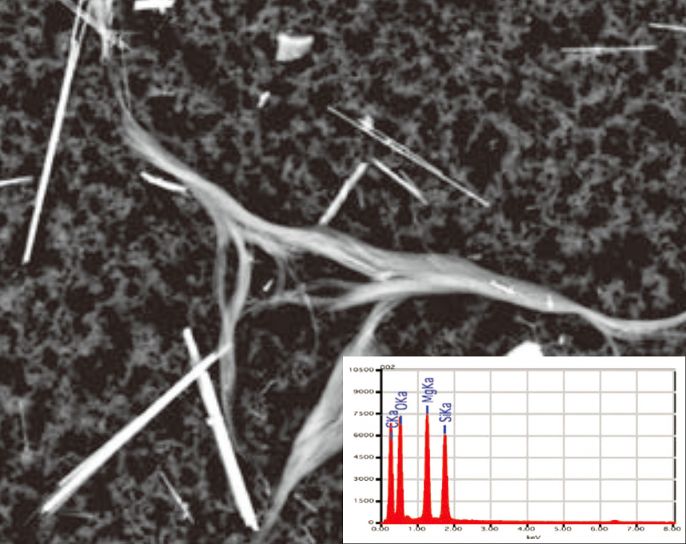 Identification of a chrysotile fibre by means of SEM imaging and EDX spectrum
Identification of a chrysotile fibre by means of SEM imaging and EDX spectrum
Bildquelle: JED-Broschüre
- Characterising layer defects
Characterising layer defects
Modern paintwork is usually a multi-layer system. In the case of macroscopically visible paint defects, it is very important to be able to determine the layer in which the cause of the defect lies. JEOL preparation systems enable the simple and reproducible production of artifact-free cross-sections.
Product groups
Solutions
Earth sciences, mineral resources, energy and other resources
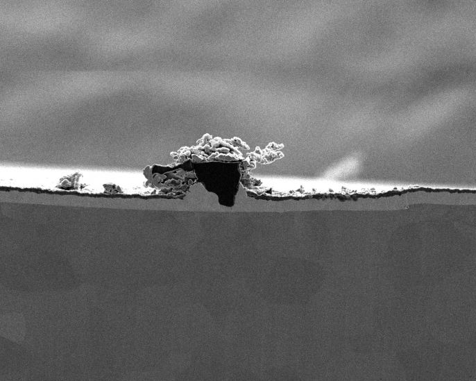 Cross-section of a painted metal surface. The diameter of the inclusion is approx. 10 µm.
Cross-section of a painted metal surface. The diameter of the inclusion is approx. 10 µm.
Source data: JEOL Ltd., CP brochure
- Characterising lithium batteries
Characterising lithium batteries
Li batteries are used in mobile phones or vehicles, among other things. Thanks to the newly developed light element spectrometer from JEOL, detecting in a microscope has for the first time become routine and standard with high spatial resolution and detection sensitivity.
Product groups
Solutions
Earth sciences, mineral resources, energy and other resources
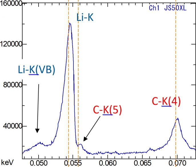 Identification of lithium in an Li-ion battery
Identification of lithium in an Li-ion battery
Source data: JEOL Ltd., SXES brochure
- Characterising precipitates
Characterising precipitates
The formation of precipitates is used systematically to define the mechanical properties of a metallic structure. However, as a form of contamination, these can also be undesired. In order to be able to judge the quality of an alloy, it is necessary to determine the morphology and chemical composition of the precipitates. This is why JEOL supplies all-round, complete solutions, from artifact-free sample preparation to high-resolution analysis from the µm to the nm level.
Product groups
Solutions
Earth sciences, mineral resources, energy and other resources
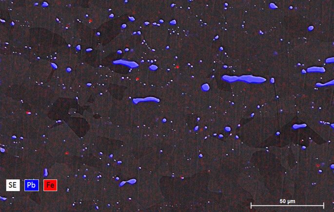 Element mapping image of a brass alloy
Element mapping image of a brass alloy
Source data: JEOL (Germany) GmbH
- Fibre analysis
Fibre analysis
Fibres are used in many branches of industry, e.g. in textile processing or as a structural material in mechanical engineering. Their structural properties can be studied by means of a fibre cross-section, for example. JEOL supplies an established and powerful complete solution for simple, artifact-free preparation and high-resolution imaging and analytics.
Product groups
Solutions
Earth sciences, mineral resources, energy and other resources
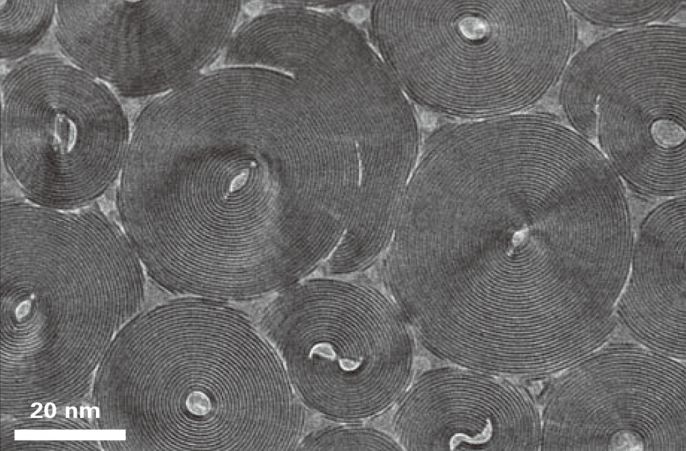 SEM image of a cross-section through a fibre bundle
SEM image of a cross-section through a fibre bundle
Source data: JEOL Ltd., Ion Slicer brochure
- Fuel analysis
Fuel analysis
Fossil fuels such as Diesel are highly complex mixtures of a wide range of linear, cyclic and aromatic hydrocarbons. Separating theses and thus unequivocally determining the constituents of the fuel is therefore of crucial importance. The combination of special, two-dimensional GC techniques allows JEOL systems to quickly deliver exact structural information.
Product groups
Solutions
Earth sciences, mineral resources, energy and other resources
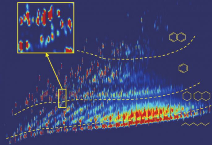 Two-dimensional total ion current chromatogram (TICC) of diesel fuel by GCxGC/TOFMS; separation after boiling point (abscissa) and polarity (ordinate)
Two-dimensional total ion current chromatogram (TICC) of diesel fuel by GCxGC/TOFMS; separation after boiling point (abscissa) and polarity (ordinate)
Source data: JEOL Ltd., JMS-T200GC AccuTOF GCx brochure, page 4
- Gas analysis
Gas analysis
The precise determination of gases with minimal differences in their atomic masses is an important task in the production of technical gases. It is not possible to exactly determine all the constituents of a gas mixture with conventional mass spectrometers. The special high-performance mass spectrometers from JEOL enable the determination and analysis of all gaseous, chemical compounds, including atomic hydrogen.
Product groups
Solutions
Earth sciences, mineral resources, energy and other resources
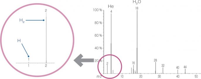 Identification of H2 and H in the mass spectrum of a gas mixture
Identification of H2 and H in the mass spectrum of a gas mixture
Source data: JEOL Ltd., JMS-MT3010HRGA brochure, page 3
- Imaging moist samples
Imaging moist samples
To achieve the high-resolution imaging and analytics of biological samples, it is often necessary to examine the sample in its native state. Thanks to the patented JEOL Aqua Cover, it is even possible to image moist or hydrated samples in a scanning electron microscope at low pressure.
Product groups
Solutions
Earth sciences, mineral resources, energy and other resources
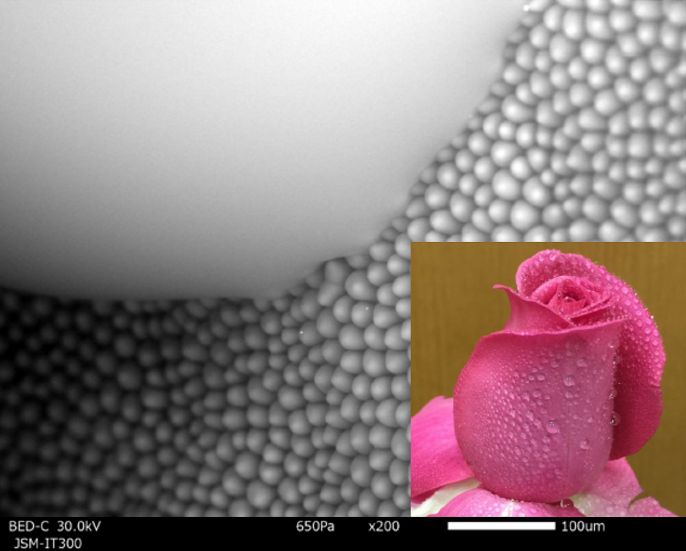 Image of a water droplet on the surface of a rose petal
Image of a water droplet on the surface of a rose petal
Source data: JEOL Ltd. (Aqua Cover presentation)
- Microscale trace element analysis
Microscale trace element analysis
The detection of rare earths is not only of interest for mineralogical samples, it is also becoming increasingly significant due to the continuous development of high-performance microelectronics. For decades, JEOL has been setting the bar for detecting the mostly low-concentrated elements with its energetically and spatially high-resolution trace element analytics.
Product groups
Solutions
Earth sciences, mineral resources, energy and other resources
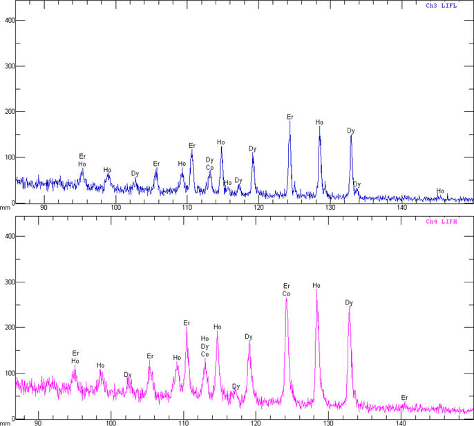 Rare earths as an example of trace element analysis
Rare earths as an example of trace element analysis
Source data: JEOL (Germany) GmbH, Demoreport Uni. Vienna (JXA)
- Mineral microstructures
Mineral microstructures
Minerals are frequently complex structures formed from a multitude of elements. Element mapping images are one of the most important methods for achieving the spatially resolved visualisation of the chemical composition. These mapping images can be used to gather essential information on e.g. the creation and structure of the samples under examination. For this task, JEOL supplies the most stable and most energetically and spatially high-resolution spectroscopy systems.
Product groups
Solutions
Earth sciences, mineral resources, energy and other resources
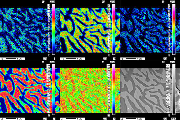 Element mapping images of a symplectite microsection
Element mapping images of a symplectite microsection
Source data: JEOL (Germany) GmbH, Demoreport Uni. Vienna (JXA)
- Petroleum analysis
Petroleum analysis
As a widespread fuel, petroleum comprises a mixture of liquid hydrocarbons. It is very difficult to perform molecular analysis on it with standard mass spectrometric methods. JEOL systems are capable of determining the molecular weight without fragmentation in a quick, uncomplicated manner by soft ionisation.
Product groups
Solutions
Earth sciences, mineral resources, energy and other resources
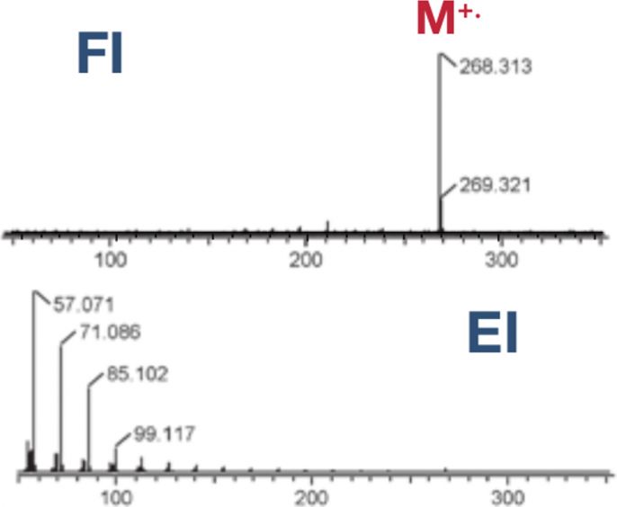 Mass spectra of an n-alkane, imaged with field ionisation (top) and electron impact ionisation (bottom)
Mass spectra of an n-alkane, imaged with field ionisation (top) and electron impact ionisation (bottom)
Source data: JEOL Ltd., 200GC-Petroleum_and_Petrochemical_Solutions brochure
- Rock analysis
Rock analysis
High-resolution element maps are of central significance for understanding geological processes. Over many decades, JEOL electron probe microanalyzers have proven to be the perfect tool for the reliable, automated examination of diffusion profiles and the spatial distribution of trace elements. Our detection threshold for this non-destructive method is less than 20 ppm.
Product groups
Solutions
Earth sciences, mineral resources, energy and other resources
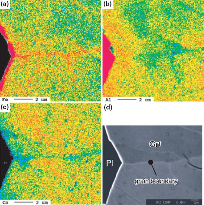 Element mapping images of a rock sample
Element mapping images of a rock sample
Source data: JEOL (Germany) GmbH, Geoforschungszentrum Potsdam
- Semiconductor analysis
Semiconductor analysis
In modern semiconductor components, complex, functional structures have to be installed in an ever smaller space. In order to be able to reliably pinpoint and identify faults, it is essential to analyse precisely the structure and the element map. With the automated systems from JEOL, it is possible to prepare, image and analyse semiconductor components for faults precisely with the greatest accuracy.
Product groups
Solutions
Earth sciences, mineral resources, energy and other resources
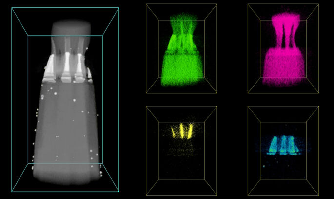 Three-dimensional element map of a NAND circuit
Three-dimensional element map of a NAND circuit
Source data: JEOL Ltd., JEM-2800 brochure/presentation
- Surface characterisation of functional materials
Surface characterisation of functional materials
More and more frequently, innovative materials are being tailor-made to their areas of application. Such precise modifications frequently occur even down to the nanoscale. Characterising such sensitive surfaces places the highest of demands on the imaging device. The high-resolution scanning electron microscopes from JEOL routinely operate within this threshold range.
Product groups
Solutions
Earth sciences, mineral resources, energy and other resources
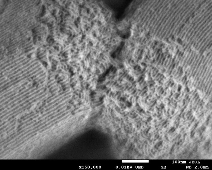 Surface image of a zeolite compound
Surface image of a zeolite compound
Source: JEOL Ltd.
